
Figure 1.
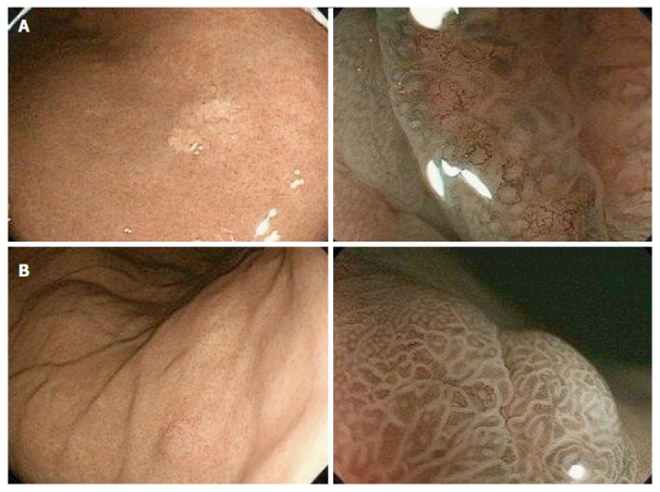
Endoscopic findings from representative GA-FG-CCP cases. A: White light endoscopy revealed a submucosal tumor-like elevated tumor with a whitish mucosal surface and dilatation of microvessels. The surrounding mucosa had no atrophic changes (left). Narrow-band imaging with magnification showed an absent microsurface pattern and irregular microvascular pattern on a small portion of the tumor (right); B: White light endoscopy revealed a submucosal tumor-like elevated tumor with normal-colored mucosal surface. The surrounding mucosa had no atrophic changes (left). Narrow-band imaging with magnification showed a regular microsurface pattern and microvascular pattern on the entire tumor surface (right).