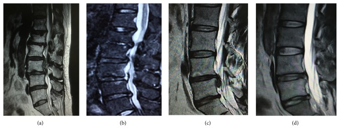
Figure 2.
Preoperative and postoperative imaging examination. (a) Preoperative magnetic resonance imaging (MRI) revealed L4/5 disc prolapse with nucleus shifting upward to L3/4 intervertebral space. (b) Postoperative MRI examination revealed clean removal of the nucleus pulposus, with no compression of the L4/5 nerve root. (c) Preoperative magnetic resonance imaging (MRI) revealed L4/5 disc prolapse with nucleus shifting downward to L5 vertebral posterior. (d) Postoperative MRI examination revealed clean removal of the nucleus pulposus, with no compression of the L4/5 nerve root.

