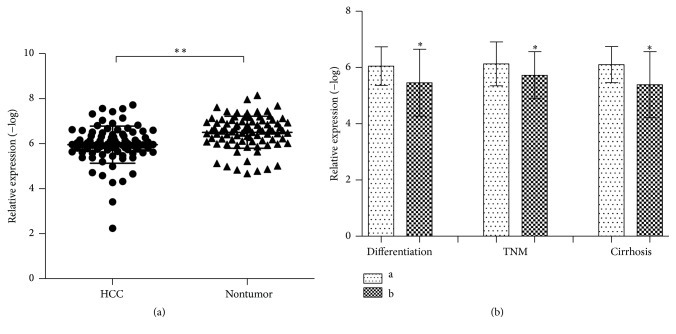
Figure 1.
Increased HERV-K (HML-2) expression in HCC tissues and the association with clinical parameters of HCC patients. The value of relative expression was detected by the value of −log of 2−ΔCt. (a) HERV-K (HML-2) expression level in tumor tissues was significantly higher than in nontumor tissues, P < 0.0001. (b) Differentiation: a: high/moderate; b: low; TNM stage: a: I-II; b: III-IV; Cirrhosis: negative/positive. Results are expressed as mean ± SD. All data were analyzed using Student's t-test. ∗ P < 0.05 and ∗∗ P < 0.01.

