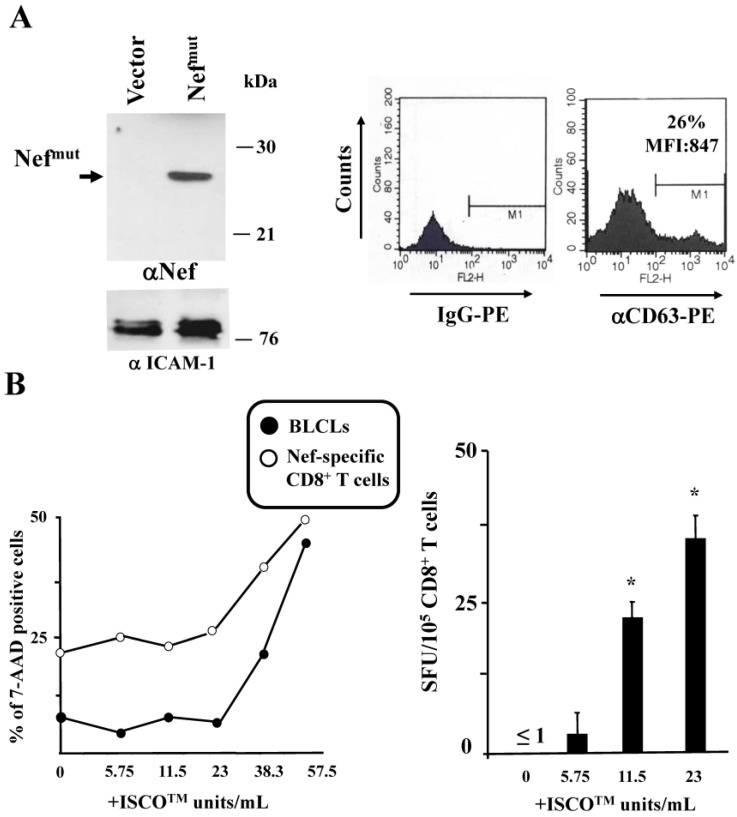
Figure 3.
ISCOMATRIXTM adjuvant increases the cross-presentation of antigens delivered by engineered exosomes in B-lymphoblastoid cell lines (LCLs). (A) molecular characterization of exosome preparations uploading Nefmut. On the left is the Western blot analysis of 200 µU of exosomes uploading Nefmut. As a control, equivalent amounts of exosomes from cells transfected with the empty vector were loaded. Arrow signs indicate the relevant protein product. Exosome preparations were also probed for the presence of Intercellular adhesion molecule (ICAM)-1, i.e., an exosome marker. Molecular markers are given in kilodaltons (kDa). On the right, fluorescence-activated cell sorting (FACS) analysis for the presence of CD63 in exosome membrane uploading Nefmu is shown. M1 marks the range of fluorescence positivity as determined by the analysis of equivalent amounts of exosomes after incubation with isotype-specific IgGs (left histogram). Both percentages and MFI of fluorescent beads are indicated. Results shown in both panels are representative of three assays carried out on two exosome preparations; (B) on the left is cell viability of both B-LCLs and Nef-specific CD8+ T cells treated for 24 h with the indicated concentrations of ISCOMATRIXTM adjuvant as evaluated by FACS analysis after 7-AAD labeling. Shown are mean values from two independent experiments with duplicates. On the right, data from cross-presentation analysis of Nefmut delivered by exosomes in B-LCLs are presented. A total of 105 cells were challenged with 100 µU of exosomes and then cultivated for 5 h in the presence of the indicated doses of ISCOMATRIXTM adjuvant. Thereafter, the cells were put in co-culture overnight with a Nef-specific, HLA-B7 restricted CD8+ T cell line in IFN-γ Elispot microwells. Shown are the mean + SD number of SFU/105 cells calculated from five independent experiments. * p < 0.05.

