Figure 5.
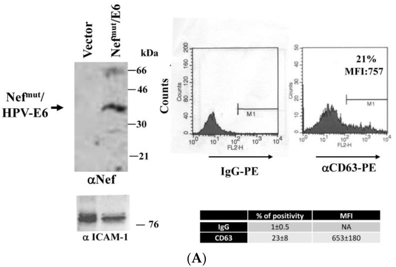
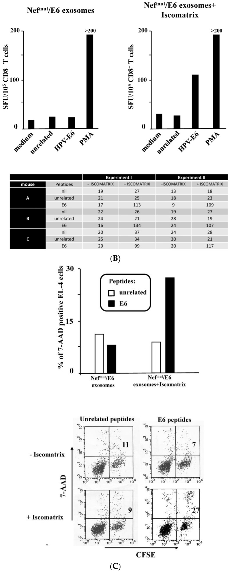
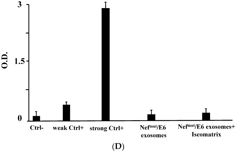
The co-inoculation in mice of ISCOMATRIXTM adjuvant and engineered exosomes increases the number of HPV-E6 specific CD8+ T lymphocytes. (A) molecular characterization of exosome preparations uploading Nefmu/HPV-E6. On the left is the Western blot analysis of 200 µU of exosomes associating Nefmut/E6. As a control, equivalent amounts of exosomes from cells transfected with the empty vector were loaded. Arrow signs indicate the relevant protein product. Exosome preparations were also probed for the presence of ICAM-1. Molecular markers are given in kDa. On the right, the FACS analysis for the presence of CD63 in exosome membrane is shown. M1 marks the range of fluorescence positivity as determined by the analysis of equivalent amounts of exosomes after incubation with isotype-specific IgGs (left histogram). Results shown in both panels are representative of five assays carried out on three exosome preparations. At the bottom right, mean values ±SD of both percentages of positivity and MFI from the five assays are also reported; (B) CD8+ T cell immune response in mice inoculated with Nefmut/E6 exosomes in the presence or not of ISCOMATRIXTM adjuvant. C57 Bl/6 mice (three per group) were inoculated s.c. at the lower right flank three times with exosomes uploading Nefmut/E6 in the presence or not of the adjuvant. Two weeks after the last inoculation, splenocytes were isolated and 105 cells were incubated overnight with or without 5 µg/mL of either unrelated or HPV-E6-specific peptides in IFN-γ Elispot microwells in triplicate conditions. As a control, untreated cells were incubated with 10 ng/mL of phorbol-12-myristate-13-acetate (PMA) and 500 ng/mL of ionomycin. Afterwards, cell activation extents were evaluated by IFN-γ Elispot assay carried out in triplicate with 105 cells/well. Cultures of splenocytes from each inoculated mouse were carried out separately. The mean of SFU/105 cells calculated on the basis of data reported at the bottom which were obtained in two independent immunization experiments are shown. Iscomatrix: ISCOMATRIXTM adjuvant; (C) Cytotoxic T lymphocyte (CTL) assay carried out with CD8+ T cells isolated from splenocytes of mice inoculated with exosomes uploading Nefmut/E6 in the presence or not of ISCOMATRIXTM adjuvant. CD8+ T lymphocytes from pooled splenocyte cultures were co-cultivated for 6 h at 20:1 effector/target cell ratio with EL (European lymphoblast)-4 cells previously labeled with Carboxyfluorescein succinimidyl ester (CFSE), and treated with either unrelated or E6 peptides for 16 h. Finally, the EL-4 cell mortality levels were scored by FACS analysis upon 7-AAD labeling. At the top, are the results obtained using pooled splenocytes from a representative of two independent immunization experiments. At the bottom, the dot-plot FACS analysis of the co-cultures is reported. Cells were gated on the basis of their apparently unaffected morphology, and the percentages of double-positive over the total of CFSE-labelled cells are reported; (D) anti-E6 antibody detection in plasma from mice inoculated with the Nefmut/E6 exosomes in the presence or not of ISCOMATRIXTM adjuvant. Plasma were tested in an in-house Elisa assay upon 1:10 dilution. As internal standards, 1:10 diluted plasma from mock-inoculated mice was used as negative control (Ctrl−), whereas both 1:1000 (weak Ctrl+) and 1:100 (strong Ctrl+) dilutions of plasma from mice injected with 10 µg of recombinant E6 protein plus adjuvant were used as positive controls. Shown are the mean absorbance values +SD of triplicates of plasma samples from each inoculated mouse.


