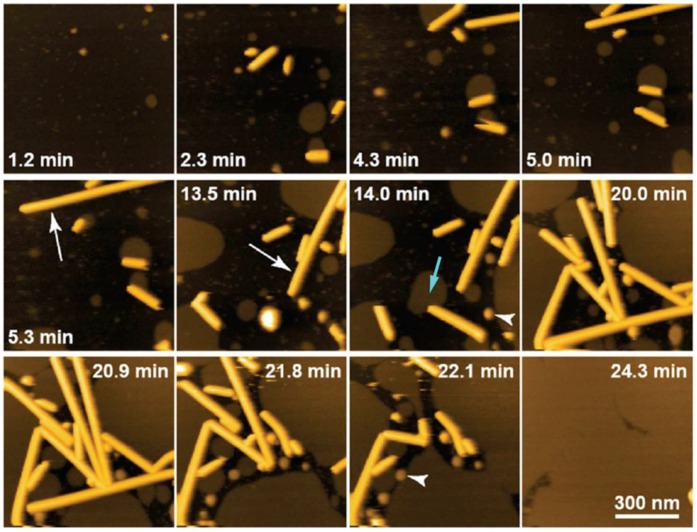
Figure 2.
HS-AFM imaging of the growth of lipid nanotubes of about 20 nm height occurring in the process of SLB formation on mica. The white arrows indicate rapidly growing lipid nanotubes. The light-blue arrow indicates the interaction between an SLB patch and one end of a lipid nanotube. The arrowheads indicate liposomes. Adapted with permission from [41]. Copyright 2014 American Chemical Society.

