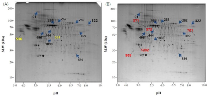
Figure 3.
2-DE gel images of the exosome proteome from U-87MG cells. (A) Exosomal proteins from U-87MG cells for 2-DE, which were incubated at 37 °C, 5% CO2 for 4 days without any change in temperature; (B) During the incubation at 37 °C, 5% CO2 for 4 days, the cells were exposed to 18 °C three times for 30 min, over a period of 36 h in a low temperature incubator (L.T.). Location of significant protein spots on 2-DE gels was represented as arrows. (blue: Spots that exist on both the control and the L.T. gel but express in different intensity; yellow: Spots that exist only on the control gel; red: Spots that exist only on the L.T. gel).

