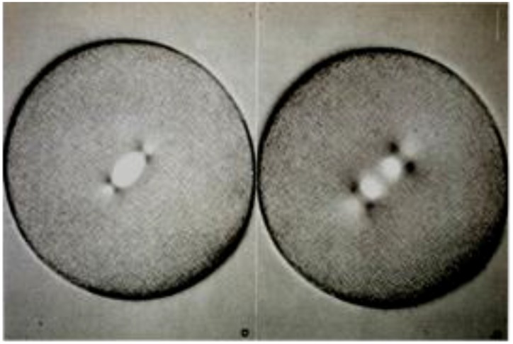
Figure 6.
Mitotic spindles in living sea urchin eggs: metaphase (left) and mid-anaphase (right) viewed with polarization microscopy, similar to Inoue and Dan, 1951 [26]. Image from Salmon, E.D., 1982, Meth. Cell Biol. 25: 69–105. With permission from the author and the Copyright Clearance Center.

