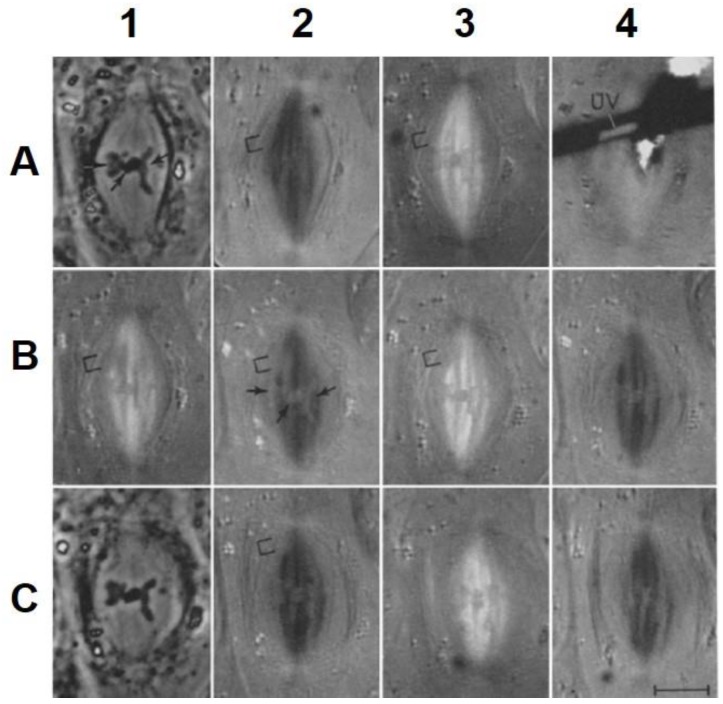
Figure 19.
A crane fly spermatocyte irradiated during metaphase. (A1) autosomes labeled with arrow; (A2,A3) the position to be irradiated is indicated by a bracket; (A4) UV = the ultraviolet irradiation; (B1,B2,B3,C2) The position of the area of reduced birefringence (on the chromosomal fiber of the left bivalent) is indicated by a bracket; (B2) the autosomes labeled with arrows. The times of the photographs in minutes relative to the time of irradiation. A1, −11; A2, −7; A3, −5; A4, −0.5; B1, +2; B2, +2.5; B3, +6; B4, +7; C1, +10; C2, +14.5; C3, +18.5; C4, +19.5. The area of reduced birefringence moved to the pole, and did not displace the pole when it arrived there. From Forer, 1966 [144]. This image is displayed under the terms of a Creative Commons License (Attribution-Noncommercial-Share Alike 3.0 Unported license, as described at http://creativecommons.org/licenses/by-nc-sa/3.0/.

