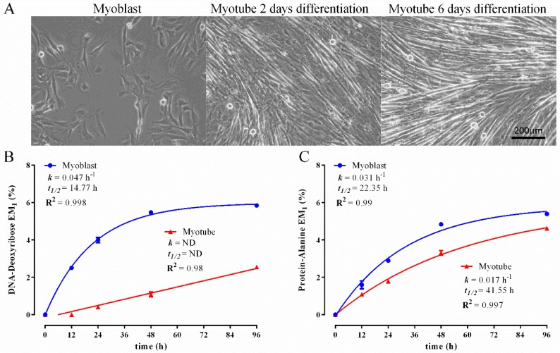
Figure 3.
Comparison of 2H-incorporation into DNA-derived deoxyribose and protein-derived alanine in C2C12 myoblasts and myotubes following incubation in 4% 2H2O treatment over 96 h. Representative images of myoblast and differentiating myotubes (A); excess molar enrichment in the M1 isotopomer (EM1) over time in DNA-derived deoxyribose (B) and protein-derived alanine (C) in myoblasts and myotubes. Inset: fractional synthesis rate constant (k), half-life (t1/2) and goodness-of-fit (R2) from non-linear curve fitting. Two replicates were performed for each time point. Error bars represent the standard error of the mean (SEM).

