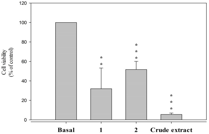
Figure 3.
Secosterols 1 and 2 decreased viability of HSC-T6 in 10 μM for 24 h. Cells were treated with DMSO (control) and coral crude extract in 6 μg/mL. Cytotoxicity assay was monitored spectrophotometrically at 450 nm. Quantitative data are expressed as the mean ± S.E.M. (n = 3–4). ** p < 0.01, *** p < 0.001 compared to basal.

