Figure 2.

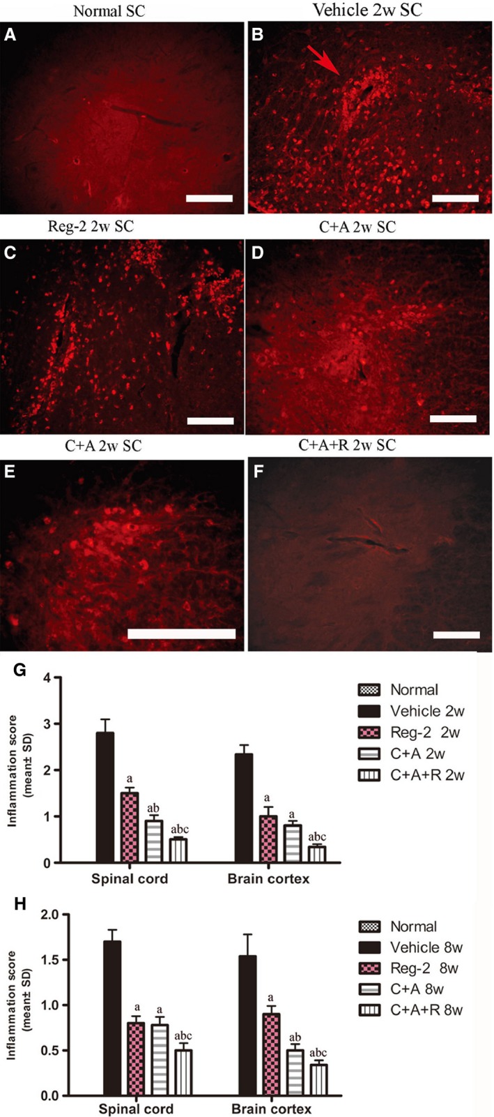
At 2 weeks Pi, infiltration of inflammatory CD68+ (a marker for extravasated macrophages) cells (B) was observed surrounding blood vessels and in the parenchyma of spinal cord in vehicle‐treated EAE rats. The C+A and Reg‐2, and especially the C+A+R, applications could evidently alleviate this phenomenon. CD68 immunofluorescence staining, scale bar: 100 μm. (E) is the regional magnification of (D), which showed the morphological feature of macrophages. The arrow in (B) shows the infiltrated inflammatory cells surrounding blood vessels as ‘perivascular cuffing’. SC, transverse sections through the anterior horn of the lumbar spinal; BC, coronal sections of the motor cortex. (G,H) C16, Ang1 and Reg‐2 treatment attenuated CNS inflammation at (G) 2 and (H) 8 weeks Pi, as shown by inflammation scoring. (a) P < 0.05 vs. vehicle‐treated EAE rats; (b) P < 0.05 vs. Reg‐2‐treated EAE rats; (c) P < 0.05 vs. C+A‐treated EAE rats.
