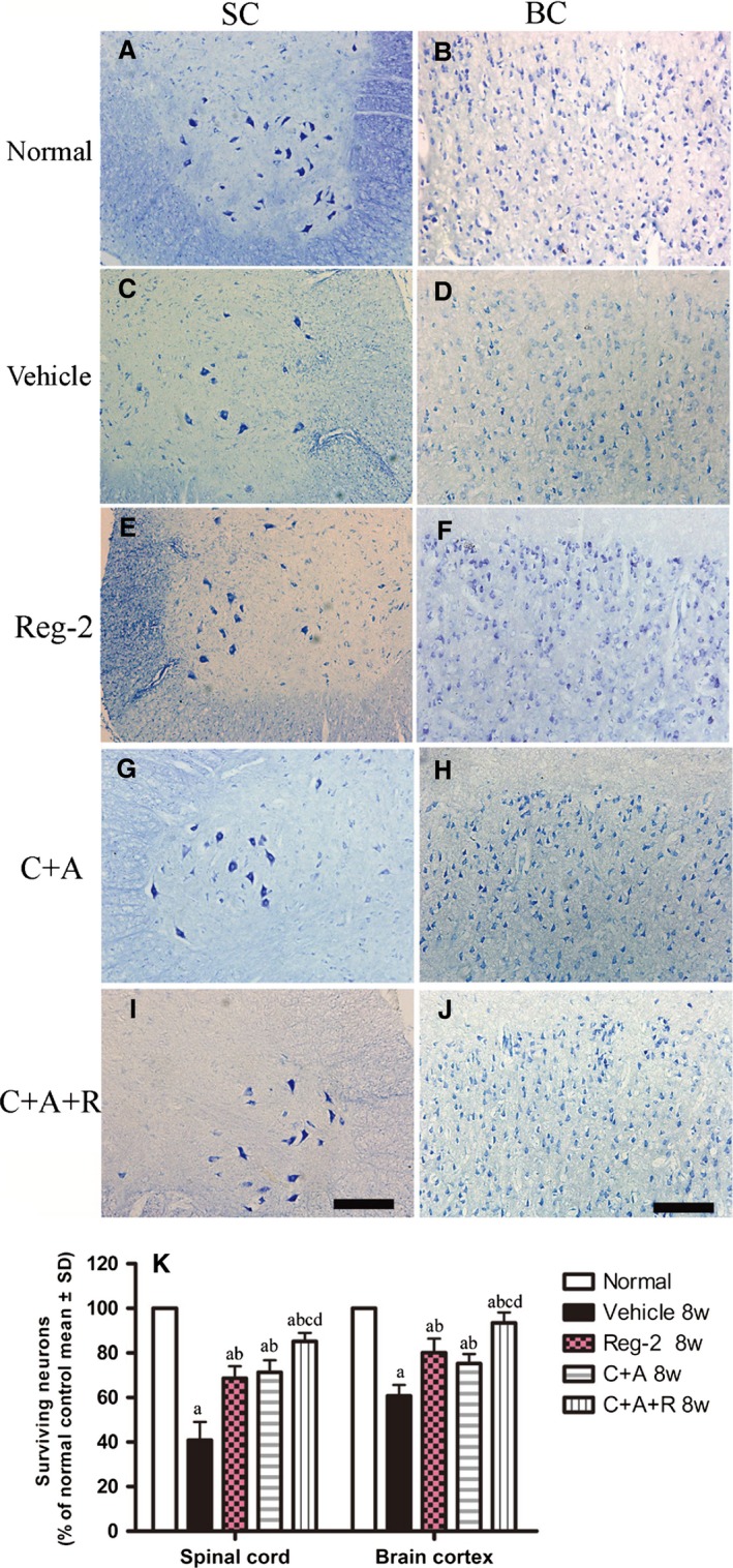
Figure 11.

(A–J) Treatment with C+A, Reg‐2 and C+A+R reduced neuronal loss in the spinal cord and brain as verified by Nissl staining at 8 weeks Pi; scale bar: 100 μm. SC, transverse sections through the anterior horn of the lumbar spinal; BC, coronal sections of the motor cortex. (K) Surviving neural cells calculated in different groups at 8 weeks Pi following Nissl staining (each group is presented as a percentage of the normal control). (a) P < 0.05 vs. normal control; (b) P < 0.05 vs. vehicle‐treated EAE rats.
