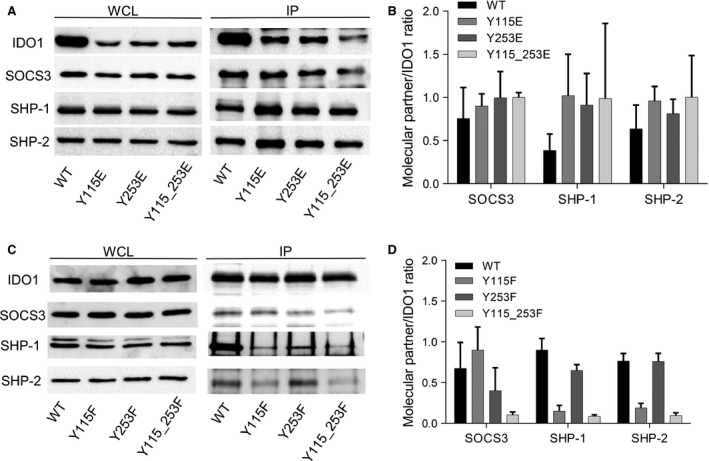
Figure 3.

Association of ITIM‐mutated IDO1 with its molecular partners. (A, C) Immunoprecipitation (IP; right panel) of IDO1 from WCL of WT and ITIM‐mutated IDO1 transfectants, and detection of IDO1 or its molecular partners (SOCS3, SHP‐1 and SHP‐2) by sequential immunoblotting with specific antibodies. WCL (left panel), immunoblot analysis of whole‐cell lysates from stably transfected cells, as control of protein expression. One representative experiment is shown. (B, D) Quantitative analysis of triplicate gels (mean ± S.D.), one of which represented in (A, C). For each cell type, the ratio of co‐immunoprecipitated protein over immunoprecipitated IDO1 was evaluated.
