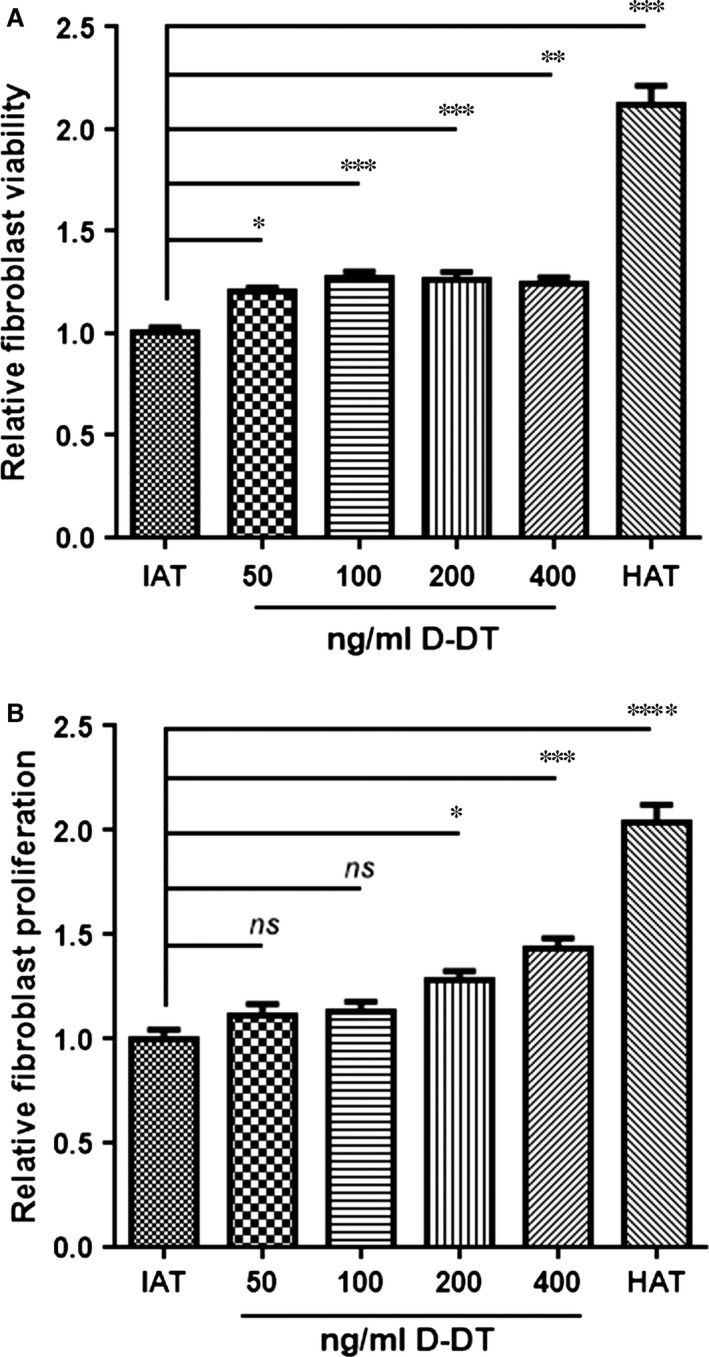
Figure 2.

In vitro viability and proliferation assay of HDF. Viability and proliferation were assessed for HDF cells. And for both assays, the cells were incubated with supernatants from HAT and IAT supplemented with varying concentrations of recombinant human D‐DT for 24 hrs. (A) Viability of HDF was measured by the alamarBlue® assay after 24 hrs stimulation with HAT or IAT supernatants supplemented with D‐DT. Cell viability increased in a dose‐dependent manner, once cells were treated with D‐DT. Graphs represent mean ± S.E.M., n = 14–16 from three independent experiments; one‐way anova with Bonferroni's multiple comparison post‐test. (B) HDF proliferation was analysed by the CytoSelect™ assay after 24 hrs stimulation with HAT or IAT supernatants with recombinant human D‐DT. D‐DT showed an induction of HDF proliferation in a dose‐dependent manner. Graphs represent mean ± S.E.M., n = 7 from two independent experiments; one‐way anova with Tukey's multiple comparisons test (*P < 0.05, **P < 0.01, ***P < 0.001, ****P < 0.0001).
