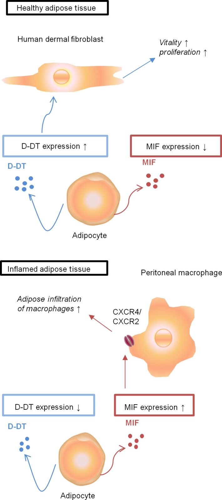
Figure 7.

Schematic illustration of the function of D‐DT and MIF in adipose tissue. Our data suggest that within healthy adipose tissue, D‐DT may increase the proliferation and vitality of human dermal fibroblasts. Upon inflammation the levels of D‐DT decrease, while MIF expression is enhanced, which subsequently may lead to an increase in peritoneal macrophage infiltration through MIF/CXCR4 or MIF/CXCR2 signalling, suggesting a pathological role for MIF in adipose tissue.
