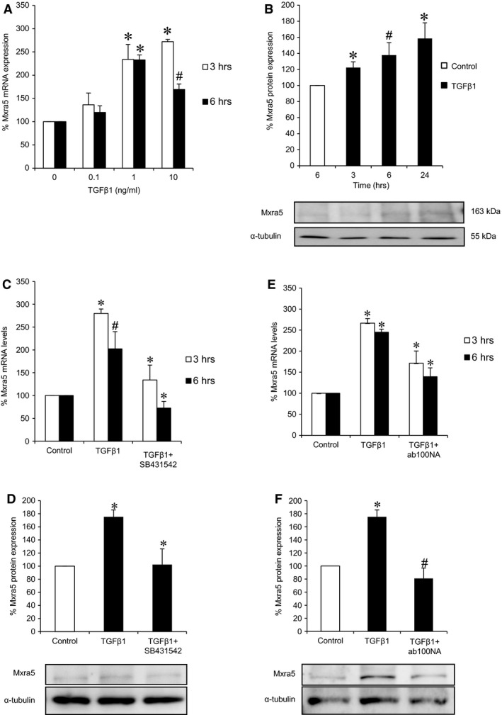
Figure 3.

TGFβ1 increases MXRA5 in cultured proximal tubular cells. (A) Human proximal tubular cells were exposed to 0.1, 1 and 10 ng/ml TGFβ1 for 3 and 6 hr and MXRA5 mRNA expression was assessed by RT‐qPCR (N = 3, *P < 0.001 versus control, #P < 0.01 versus control). (B) Cells were exposed to 1 ng/ml TGFβ1 for 3, 6 and 24 hr and MXRA5 protein expression was assessed by Western blot. Tubulin was used as loading control (N = 3, *P < 0.025 versus control, #P < 0.05 versus control). (C) Cells were pre‐treated with 10−5 M TGFβ1 receptor 1 inhibitor SB431542 for 1 hr and then exposed to 1 ng/ml TGFβ1 for 3 and 6 hr, MXRA5 mRNA expression was assessed by RT‐qPCR (N = 3, *P < 0.001 versus control, #P < 0.006 versus control). (D) Cells were pre‐treated with 10−5 M SB431542 for 1 hr and then exposed to 1 ng/ml TGFβ1 for 6 hr, MXRA5 protein levels were assessed by Western blot (N = 3, *P < 0.005 versus control). (E) Cells were pre‐treated with 1 ng/ml neutralizing anti‐TGFβ1 antibody ab100NA for 1 hr and then exposed to 1 ng/ml TGFβ1 for 3 and 6 hr, MXRA5 mRNA expression was assessed by RT‐qPCR (N = 3, *P < 0.001 versus control). (F) Cells were pre‐treated with 1 ng/ml neutralizing anti‐TGFβ1 antibody ab100NA for 1 hr and then exposed to 1 ng/ml TGFβ1 for 6 hr, MXRA5 protein levels were assessed by Western blot (N = 3, *P < 0.005 versus control, #P < 0.018 versus control).
