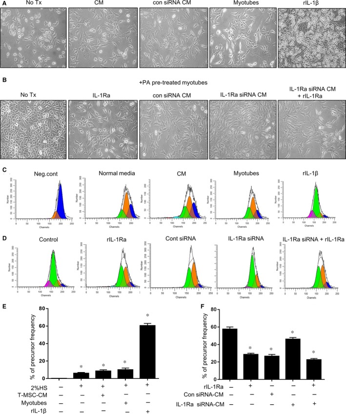
Figure 4.

Proliferation of L929 fibroblasts in the exposure to PA‐stimulated myotubes was attenuated by T‐CM containing IL‐1Ra. (A) L929 cells labelled with CFSE were seeded in the bottom of a 24‐well transwell plate. T‐CM or T‐CM from control siRNA‐transfected T‐MSCs was added to the media to test their basal activity on the proliferation of L929 cells. In the upper insert, myotubes or rIL‐1β (100 ng/ml) were added, respectively. The proliferated L929 cells were monitored using a phase‐contrast microscope after culturing for 3 days. Original magnification, 200×. (B) To confirm the effect of PA‐treated myotubes on the proliferation of L929 cells, myotubes pre‐treated with PA were placed in the upper chamber in new media. rIL‐1Ra (100 ng/ml) or T‐CM from T‐MSC transfected with either IL‐1Ra‐specific siRNA or control siRNA was supplemented to the lower chamber containing L929 cells. In addition, L929 cells in the bottom chamber were treated with a combination of rIL‐1RA (100 ng/ml) and T‐CM from T‐MSCs transfected with Il‐1ra‐specific siRNA. Microscopic observations are presented. Original magnification, 200×. (C–F) After culturing for 3 days, L929 cells in the lower chamber were collected. CFSE+ cells were measured for the degree of proliferation via flow cytometry and were analysed using the ModFit LT ™ software based on the reduction in CFSE positive cells. Result presented in (D) and (F) is from cells exposed to PA pre‐treated myotubes that located in the upper chamber. The data are presented as the mean ± SEM (*P < 0.05).
