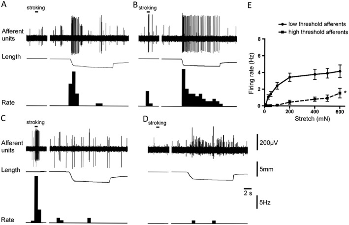
Figure 1.

Typical tracings representing responses of two groups of afferents: low threshold stretch‐sensitive afferents (A, B) and high threshold afferents (C, D) to stretch and mucosal stroking. (A) Muscular afferent (large amplitude unit) responded to small stretch (50 mN load) but not to light (0.1 mN) von Frey hair stroking of its receptive field. (B) Muscular‐urothelial afferent (large amplitude unit) was activated by stroking of its receptive field with a 0.1 mN von Frey hair and by small stretch (50 mN load). (C) Urothelial (mucosal) afferent (large amplitude unit) was activated by a light (0.1 mN) von Frey hair stroking of its receptive field area, but not by high intensity stretch (200 mN load). (D) High threshold afferent (large amplitude unit) was slightly activated by a high intensity stretch (200 mN load) but not by a light (0.1 mN) von Frey hair stroking of its receptive field. Note in each trace the largest amplitude units were discriminated and used to calculate firing rate as shown in each panel. (E) Averaged data of the effects of stretch (10–600 mN) on the low threshold stretch sensitive (shown by solid line, n = 18, N = 14) and high threshold (shown by dashed line, n = 21, N = 15) afferents. *P < 0.05, significantly different from low threshold values.
