
Figure 6.
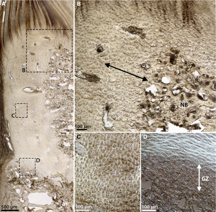
DIC images of the cells within the newborn cartilaginous endplate at (A) low magnification and (B‐D) corresponding high magnification. (B) Region adjacent to the mass of new bone tissue (NB). (C) A peripheral region deeper within the cartilage. (D) The growth plate. Note the changing cell appearance in (B) where the cells grow progressively larger and seem to align near the vertebral bone (see arrow), and in (D) where the cells in the growth zone (GZ) appear more spherical, whereas those immediately above this zone are more horizontally aligned. In (C) and through the rest of the cartilaginous tissue, however, the cells appear undistorted and more randomly arranged.