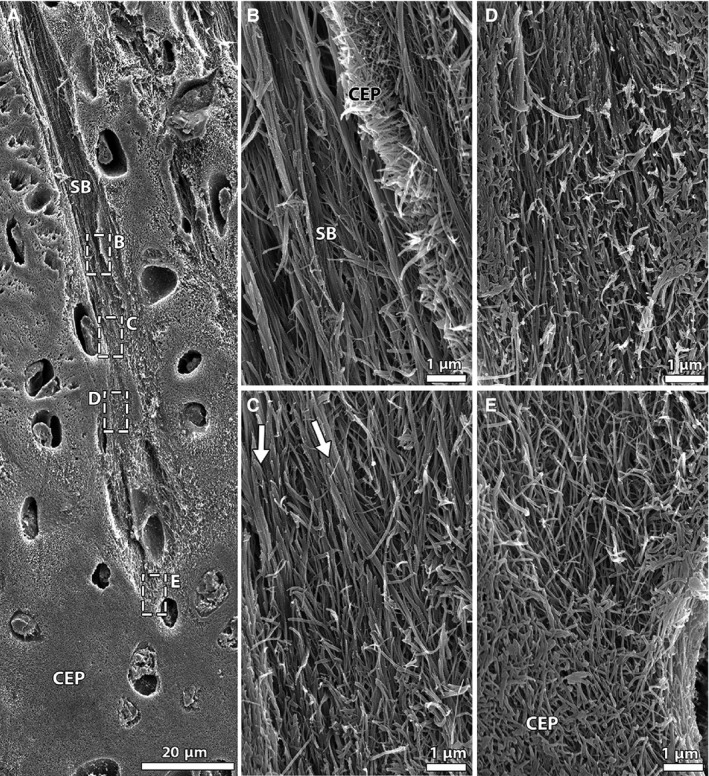
Figure 7.

SEM images capturing fibrillar integration between annular sub‐bundles and cartilaginous endplate in the newborn disc. (A) Low magnification; (B‐E) high magnification views of boxed regions in image (A) at various depths of anchorage. Note the change in fibril appearance, from thick aligned fibrils higher up within the sub‐bundle (B) to a fine network of randomly arranged fibrils at its end (E). The arrows in (C) highlight a group of sub‐bundle fibrils splitting into smaller units that merge with the surrounding fibrils. CEP, cartilaginous endplate; SB, sub‐bundle.
