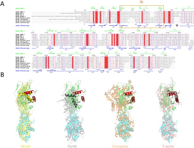
Figure 6.

Comparison with other actin‐like filaments. (A) Multiple sequence alignment of MamK and MreB orthologs from a number of bacterial strains. Identical residues are in a red box, similar residues are in red character. The secondary structure elements for MamK and MreB are shown at the top in green, and at the bottom in blue, respectively. Domain Ib is indicated with a pink box, and the loop involved in cross‐strand contacts in MamK is shown with a purple star. (B) Comparison of the filament arrangement between MamK (green and cyan) and MreB (crystal structure‐derived filament, yellow), ParM (gray), Crenactin (orange), and F‐actin (pink). For each of them, two adjacent molecules along the helical axis are shown, where the bottom subunit is aligned to the bottom (green) subunit of MamK. For clarity, two helices from domain Ia are in red in the MamK structures, and highlighted in the other bacterial actins. Despite the closer sequence and structural similarity of the MamK protomer to MreB, the top (green) subunit of MamK aligns more closely to that of F‐actin.
