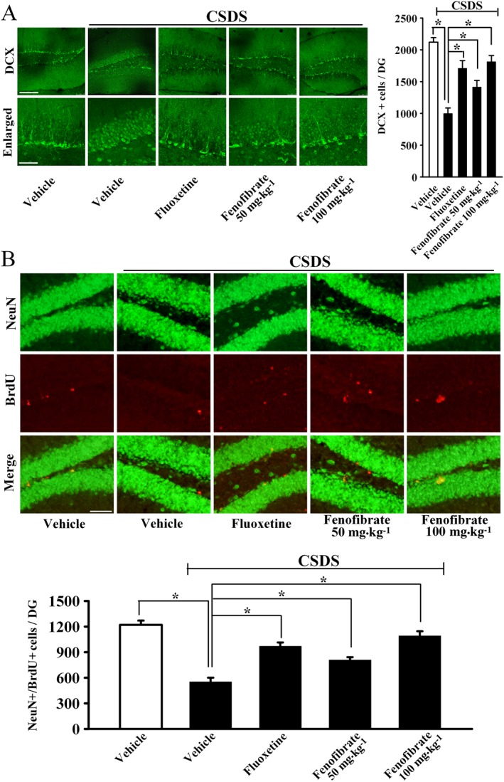
Figure 3.

Fenofibrate administration restores the decreased adult hippocampal neurogenesis caused by CSDS stress. (A) Representative confocal microscopic images showing the localization of doublecortin (DCX; green) in the dentate gyrus (DG). The scale bar is 150 μm for representative images and 50 μm for enlarged images respectively. Density statistics showed that chronic fenofibrate treatment significantly increased the number of DCX‐stained cells in the DG of stressed mice. (B) Representative microscopic images showed the co‐staining (yellow) of neuronal nuclei (NeuN) (green) and BrdU (red) in the DG. The majority of BrdU+ cells are doubly labelled with the neuronal marker NeuN and located within the granule cell layer. The scale bar is 75 μm. Density statistics showed that fenofibrate treatment fully reversed the CSDS‐induced decrease in the number of NeuN+/BrdU+ cells in the DG. Data are expressed as means ± SEM (n = 5); * P < 0.05. Comparisons were made by one‐way ANOVA followed by post hoc LSD test.
