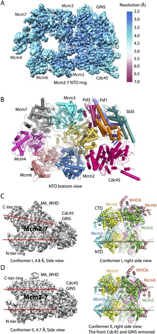
Figure 5.

Cryo‐EM structure of the yeast CMG helicase. A: 3D cryo‐EM map of CMG in the bottom N‐terminal view color coded by local resolution (EMD ID: 6534). B: CMG atomic model viewed from the N‐tier face (PDB ID: 3JC6). C: CMG conformer I at 4.8 Å resolution (EMD ID: 6536, PDB ID: 3JC6). D: CMG conformer II at 4.7 Å resolution (EMD ID: 6535; PDB ID: 3JC5). The two red or black lines in C, D: highlight the distinct configuration of the two conformers. The dashed blue line encircles Mcm2 AAA+ domain. This domain is rotated by 30° in conformer II. Note that the right panels in (C, D) are rotated by 90° with respect to the left panels.
