Fig. 2.
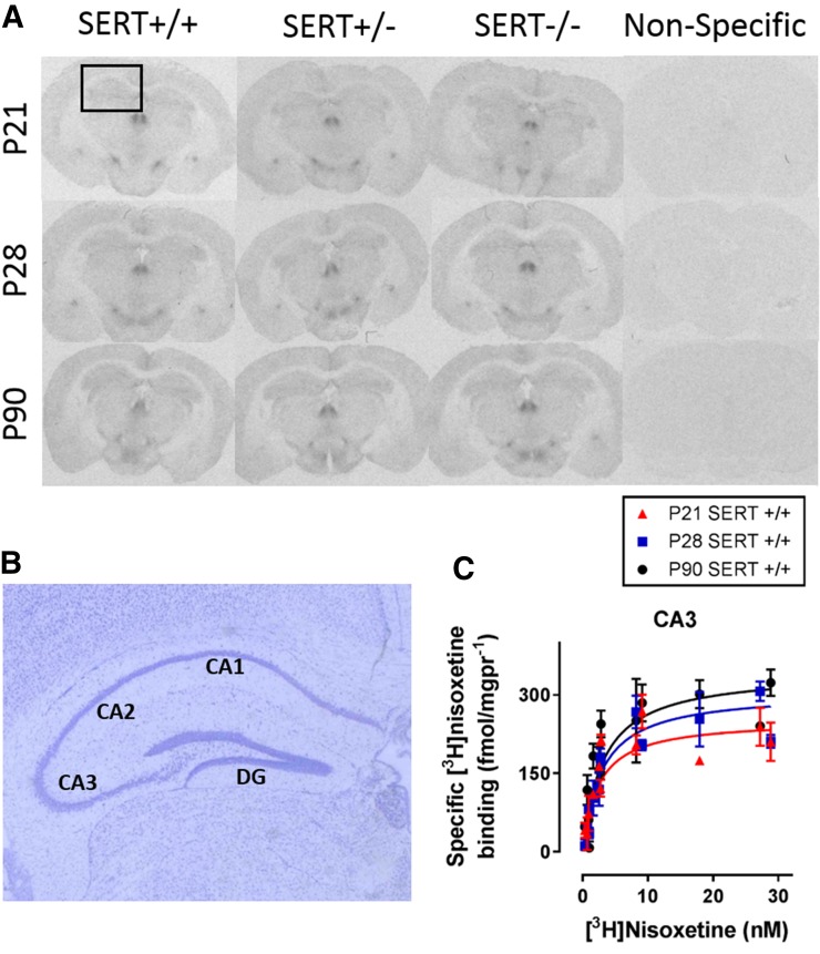
Specific [3H]nisoxetine binding to NET in hippocampal regions as a function of age and SERT genotype. Brain sections from P21, P28, and P90 SERT-deficient mice incubated with the NET-specific ligand [3H]nisoxetine. Nonspecific binding was defined by mazindol (2.5 mM). (A) Representative coronal sections at the level of plate 47 (Paxinos and Franklin, 1997) in SERT+/+, SERT+/−, and SERT−/− mice aged P21, P28, or P90. The boxed area in (A) is enlarged in (B), which shows representative thionine-stained brain sections labeled with hippocampal regions quantified, which include the CA1, CA2, and CA3 regions and the dentate gyrus (DG). (C) Example of saturation binding isotherms used to calculate Bmax and Kd values. Curves include specific [3H]nisoxetine binding values for the CA3 of P21, P28, and P90 SERT+/+ mice. There was no main effect of sex on Bmax or Kd values, so male and female data are pooled (P > 0.05). Bmax values are summarized in Fig. 4. There were no significant differences in Kd among ages or between SERT+/+ and SERT+/− mice. Sample sizes of mice per group were as follows: SERT+/+, n = 5–9 (4 males and 3–5 females, pooled); SERT+/−, n = 6–10 (3–5 males and 2–4 females, pooled); and SERT−/−, n = 4–7 (2–4 males and 2–4 females, pooled). See Table 2 and Fig. 4 for a summary of data.

