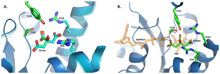
Figure 2.
Citrate binding in the 2-Kase domain. (A) Superimposed structures of the citrate binding pocket for human (blue) and bovine (light gray) orthologues. Residues forming interactions with citrate are represented as sticks, with a ribbon diagram representing the mainchain. Human PFKFB2 is depicted as a blue ribbon with sticks containing green carbon atoms. In contrast, bovine PFKFB2 is represented as a light gray ribbon with sticks containing white carbon atoms. (B) AMPPCP and F6P from PFKFB3 (2DWP) overlaid onto the citrate binding site of human PFKFB2. The distance between the carboxy arm from citrate and nearest atom from the overlaid AMPPCP is shown. Both AMPPCP and F6P are semi-transparent colored orange.

