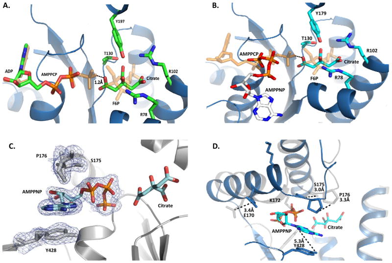
Figure 3.
Ligand Binding within the 2-Kase Domain. (A) Bovine PFKFB2 complexed with ADP and Citrate. Superimposed are AMPPCP and F6P from the pseudo-substrate complex with PFKFB3 (2DWP), showing the catalytic ATP and F6P binding modes, respectively. (B) Human PFKFB2 complexed with AMPPNP and Citrate. Superimposed are AMPPCP and F6P from the pseudo-substrate complex with PFKFB3 (2DWP), showing the catalytic ATP and F6P binding modes, respectively. (C) Unbiased Fo-Fc omit map of AMPPNP and neighboring residues in human PFKFB2. Contoured at 3.0σ (D) Ribbon diagram view of human PFKFB2 (blue) super-imposed onto the bovine orthologue (light gray). The sidechains and their positional differences are shown for residues involved in AMPPNP binding.

