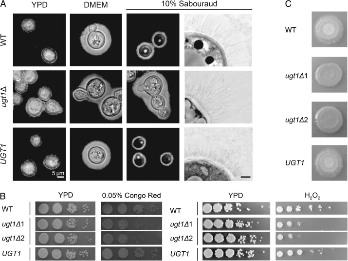
Fig. 6.
ugt1Δ mutants show altered capsule and cellular morphology, and exhibit growth and mating defects. (A) Wild-type (WT), ugt1Δ and complemented ugt1Δ (UGT1) were grown in the media noted above and visualized by light microscopy after negative staining with India Ink (first three columns, scale bar = 5 μm) or by electron microscopy (last column, scale bar = 500 nm). (B) The indicated strains, including two independent ugt1Δ strains, were grown overnight at 30°C in YPD, and 5 μl of serial dilutions were spotted and grown as indicated. Left panel, dilutions were 10-fold starting at 106 cells; right panel, dilutions were 5-fold, starting at 107 cells. (C) Equal volumes of the indicated MATα strains and KN99a were mixed, spotted on V8 agar and incubated at RT in the dark. Images were taken 2 weeks after initial spotting. In three independent experiments, no filamentation of either mutant strain was detected.

