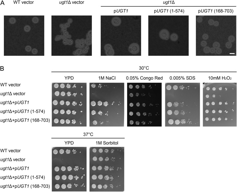
Fig. 7.
N- and C-terminal UGT1 truncations complement capsule and cellular morphology defects in ugt1Δ. (A) The strains indicated above the horizontal line, carrying the plasmids shown below the line, were grown in DMEM + G418 and visualized by light microscopy after negative staining with India Ink (scale bar = 5 μm). (B) The indicated strain and plasmid combinations were grown overnight at 30°C in YPD + G418. The 5 μl of serial dilutions were spotted and grown as indicated with G418 (except for 0.005% SDS). Dilutions were 5-fold starting at 106 cells.

