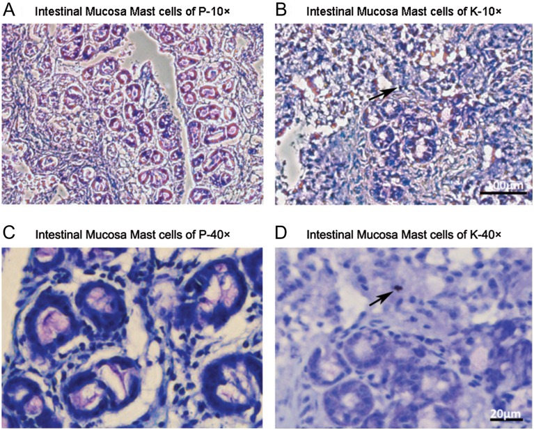
Fig. 4.
Micrographs of intestinal mucosa of piglets stained with toluidine blue. Mast cells are stained violet color with blue background (details in methods and material). (A and B) Intestinal sections of P and K, respectively, at 10× (Bar = 100 μm). (C and D) Intestinal sections of P and K, respectively, at 40× (Bar = 20 μm). Mast cells were not found in the P samples, while only one mast cell, indicated with black arrows, was present in the K samples. n = 4–5 sections per sample. This figure is available in black and white in print and in color at Glycobiology online.

