Figure 5.
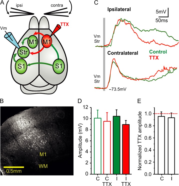
Blocking contralateral M1 does not affect striatal response to ipsilateral whisker stimulation. (A) Diagram of the experimental configuration and main synaptic pathways; arrows in red show the synaptic outputs blocked by TTX injection to contralateral M1. Unblocked regions and pathways are shown in green, the recording electrode in dorsolateral striatum in blue, and the TTX injection in M1 of the contralateral cortical hemisphere marked in red (see also Fig. S1). (B) Example of a TTX (10 µM) and neurobiotin (0.4%) injection-site in contralateral M1. (C) Waveform average (>40 repetitions) of a whole-cell recorded MSN in response to contra- and ipsilateral whisker deflection before and after injection of TTX 10 µM in contralateral M1. The gray bar represents the air-puff whisker deflection. (D) Response amplitudes to contralateral and ipsilateral whisker deflection (n = 14). (E) Normalized response amplitudes. The normalization was done with respect to the control condition for each neuron and type of stimulation (n = 14). All responses are measured during down states. Asterisks *, **, *** represent P values <0.05, <0.01, <0.001, respectively.

