
Figure 3. Porcupine inhibition improves cardiac function and reduces adverse remodeling after MI.
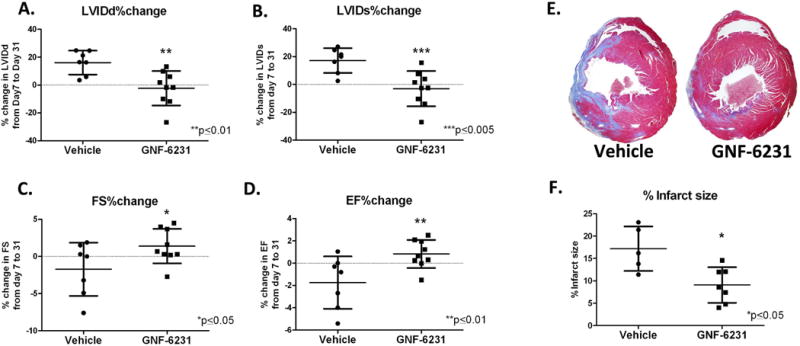
Left ventricular remodeling was measured as % change in (A) LVIDd and (B) LVIDs. LV function was measured as % change in (C) FS and (D) EF. Data showed no increase in left ventricular diameter (A and B), and improved cardiac function (C and D) with GNF-6231 treatment compared to vehicle. (E) Masson’s trichrome stained representative sections of the left ventricle at day 30 depicted more collagen stained (blue) area in vehicle-treated LV compared to GNF-6231-treated. (F) Quantification of infarct size. Each data point on graphs represents individual mouse; *p ≤ 0.05, **p ≤ 0.01 or ***p ≤ 0.005; unpaired t-test.