
Figure 5. WNT inhibition increases proliferation of progenitor cells that may contribute to myogenesis.
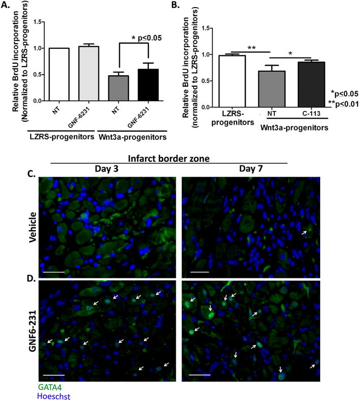
(A and B) Relative BrdU incorporation by Sca1+ progenitor cells stably expressing LZRS (empty vector) or Wnt3a-LZRS revealed that proliferation was reduced by Wnt3a overexpression and this effect was reversed by (A) GNF-6231 treatment and (B) C-113 treatment.
Data are presented as Mean ± SD. (A) N=5 and (B) N=3 replicates from independent experiments; *p ≤ 0.05 and **p ≤ 0.01; Kruskal-Wallis test with Dunns correction for multiple comparisons. (C and D) Representative GATA4 immunostained sections of infarct border zone at day 3 (left panels) and day 7 (right panels) post-MI of (C) vehicle-treated hearts and (D) GNF-6231 treated hearts. White arrows point to GATA4 stained nuclei. Scale bars equal 50 μm; the images are representative of at least 4 sections each from N ≥ 3 mice per group.