
Figure 6. WNT inhibition reduces cardiomyocyte cell death.
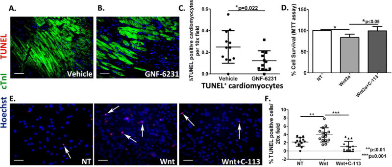
(A and B) Representative sections co-stained with cTnI and TUNEL of infarct border zone of (A) vehicle-treated hearts, and (B) GNF-6231 treated hearts. (C) Quantification of percent TUNEL positive cardiomyocytes revealed significantly fewer apoptotic cardiomyocytes in GNF-6231 treated hearts. N=5 mice per group, at least 4 sections imaged per mouse, *p=0.022, unpaired t-test. (D) Percent cell survival of HL-1 rat cardiomyocytes as measured by metabolic uptake (MTT) assay showed significant reduction in survival with recombinant WNT3A treatment, which was rescued by addition of C-113. Bars represent mean ± SD; N=3 replicates from independent experiments; *P ≤ 0.05; Kruskal- Wallis test with Dunns correction for multiple comparisons. (E) Representative images of human iPSC-derived iCell cardiomyocytes treated with recombinant WNT3A or WNT3A and C-113 in presence of 250 μM H2O2 showing that under stress, treatment with recombinant WNT3A increased cell death, which was rescued by WNT inhibition with C-113. (F) Quantification of %TUNEL positive iCell cardiomyocytes per 20× field. N ≥ 12 per group, at least 4 areas imaged in each replicate from 3 independently run experiments; **p ≤ 0.01 and ***p ≤ 0.001; One-Way ANOVA with Bonferroni correction for multiple comparisons. Scale bars in A-B and E represent 50 μm.