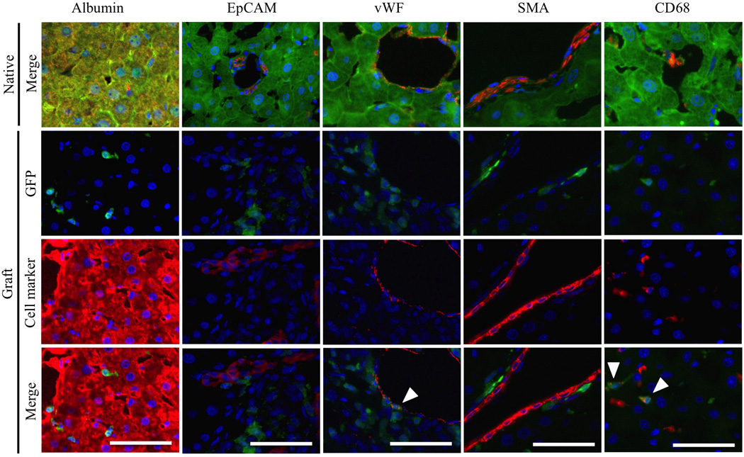
Figure 4. Detection of host-derived cells in liver grafts.

Staining was performed on native livers and liver grafts for hepatocytes (Alb), cholangiocytes (EpCAM), sinusoidal and vessel endothelial cells (vWF), hepatic stellate cells (αSMA) and Kupffer cells/monocytes (CD68), together with GFP immunofluorescence, 4 weeks after APLT. White arrowheads indicate co-localization. Scale bars = 50µm.
