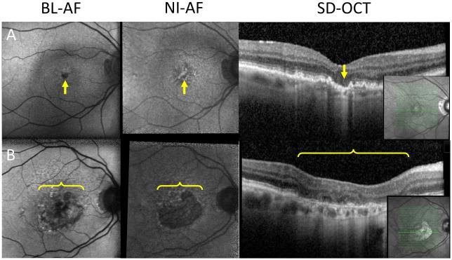
Figure 3.
Geographic atrophy missed on BL-AF.
A: Left column: geographic atrophy isolated to the fovea (arrow), which normally appears as hypo BL-AF in healthy patients. Middle column: geographic atrophy visualized on NI-AF as a hypo NI-AF lesion (arrow). Right column: OCT showing area of increased signal backscattering into the choroid indicating the absence of RPE and the presence of geographic atrophy (arrow). B: Left column: heterogeneous area of hypo BL-AF and normal BL-AF (bracket). Middle column: homogenous well demarcated area of hypo NI-AF indicating geographic atrophy (bracket). Right column: OCT indicating the presence of geographic atrophy (bracket). Inset: location of OCT section.

