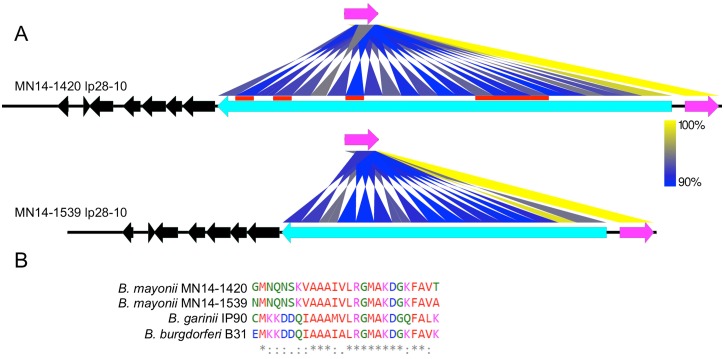
Fig 6. vls containing linear plasmid lp28-10 from B. mayonii MN14-1420 and MN14-1539.
A) Heat map displaying nucleotide identity (blue—lowest; yellow—highest) between the active vlsE nucleotide sequence (magenta arrow) and silent vls cassettes within the two B. mayonii strains. Position and copy number of the silent cassette array is indicated by the blue to yellow ribbons. Red bars indicate silent vls loci present in MN14-1420 and missing in MN14-1539. Genes flanking the vls locus are indicated by black arrows. Orientation of arrows indicates gene orientation on the forward or reverse DNA strand. B) Multi-alignment of the 26 amino acid C6 peptide within VlsE from B. mayonii, B. burgdorferi B31 and B. garinii IP90. Identical (*), conserved (:) and differentially charged (.) amino acid residues are indicated.

