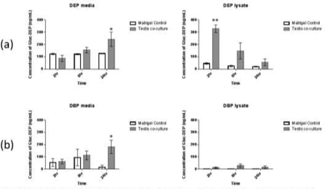
Fig. 3. Concentration of glucuronidated metabolite in testis co-culture cell media and cell lysate after treatment with phthalate esters for 24 hours.
Tests co-cultures were treated with (a) diethyl phthalate (DEP, 150µM) or (b) dibutyl phthalate (DBP, 100µM). Levels of monoester metabolites were measured before and after glucuronidase treatment in order to determine levels of glucuronidated metabolites in each sample. Bars show mean +/− s.e. (n=3) *: p<0.05, **: p<0.001 (t-test vs. corresponding matrigel control)

