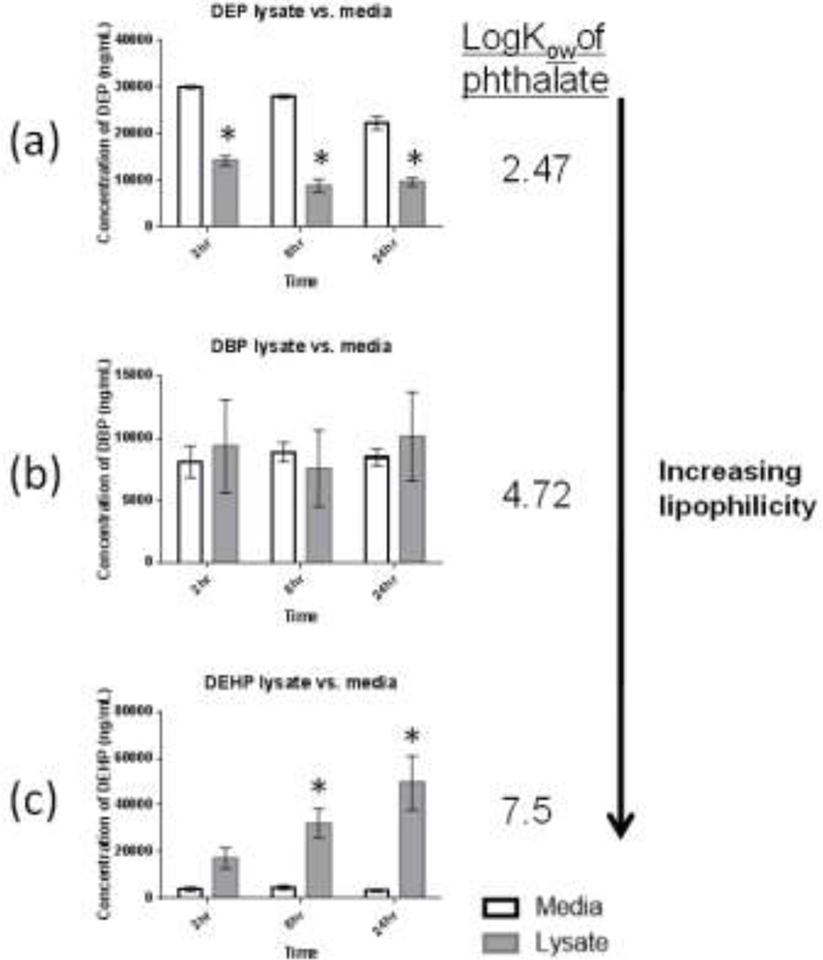
Fig. 5. Concentrations of 3 phthaiate diesters detected in testis co-culture cell media or cell lysate after exposure to either DBP, DEHP (100µM) or DEP (150µM).
Testis cells were exposed to DBP, DEHP or DEP for 2, 8 or 24 hours. Values are mean and standard errors for 3 replicate treatments. Bars show mean +/− s.e. (n=3) * indicates average concentration in cell lysate samples were significantly different then concentration in cell media at corresponding time point (t-test p<0.05).

