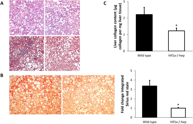
Fig 4. Liver histology and collagen quantification.
(A) Representative liver H&E (top) and Masson’s trichrome stains (bottom) from wild-type (left) and Hif1a-/-hep mice. More fibrosis can be observed in the wild-type mice in the Masson’s trichrome stain. (B) Sirius red stain of collagen in wild-type (left) and Hif1a-/-hep mice (right). (C) Collagen content of all samples by use of hydroxyproline assay. *, p<0.05.

