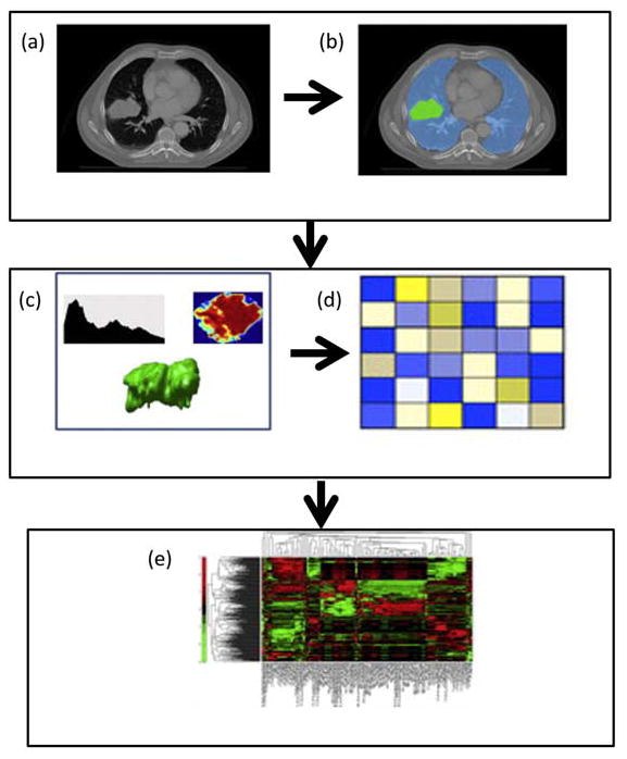
Figure 1.
Illustration of the radiomics algorithm. (a) Initial Computed Tomography (CT) scan. (b) Segmentation is performed on the lesion using a region of interest(ROI). (c) Radiomic features are extracted from the ROI based on the gray level patterns, inter-voxel relationships, and shape. (d) A subset of the extracted radiomic features is selected for classification (e) The selected features are used as inputs into a classification model to produce a diagnosis or correlation to a prognostic marker. Data from5,12.

