
Fig. 6. Post-injury administration PFT-α (O) at 5 h after TBI significantly decreased p-p53 and PUMA positive neurons in the cortical contusion region at 8 h.
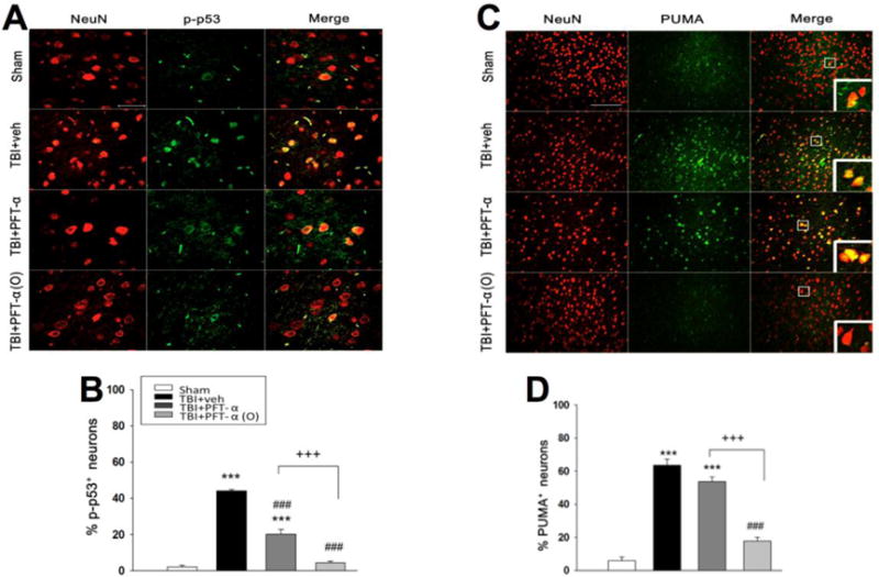
(A) Co-immunohistochemistry of p-p53 and NeuN in cortical brain tissue. (C) Co-immunohistochemistry of PUMA and NeuN in cortical brain tissue. Phospho-p53 or PUMA immunoreactivity is shown in green, and NeuN is shown in red. Yellow labelling indicates colocalization. (B, D) There was a significantly decrease in the number of p-p53 and PUMA positive neurons in TBI + PFT-α (O) group, respectively. Data represent the mean ±SEM. **p<0.01; ***p<0.001 versus the sham group; ##p<0.001; ###p<0.001 versus the TBI + veh group; ++p<0.01; +++p<0.001 versus the TBI + PFT-α group. Scale bar=100 μm. (n=5 for each group).