
Fig. 7. PFT-α and PFT-α (O) reduce glutamate-induced excitotoxicity in cortical cultures.
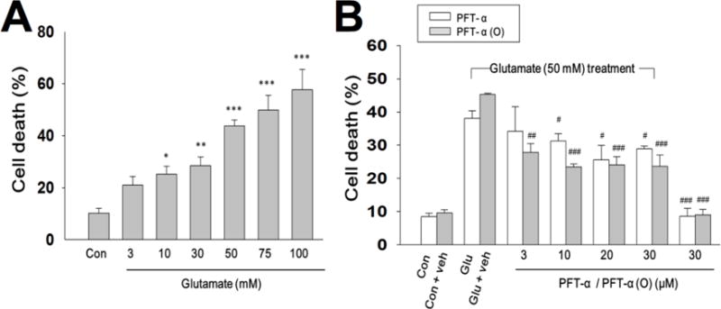
(A) Cultured cells were exposed to various concentrations of glutamate (3, 10, 50, 75 and 100 mM) for 24 h. Cell death (%) was measured by LDH release. 50 mM glutamate was chosen as the concentration for testing PFT analogues. (B) PFT- α or PFT- α (O) (3, 10, 20, 30 μM) was added 30 min after cells were exposed glutamate (50 mM) for 24 hrs in primary cultures of neuron/glia. Cell death was measured by LDH activity in culture media which was scaled to the value of maximal death (100%) measured after freeze-thaw treatment of sister cultures. Data are expressed as means±SEM. *p<0.05; **p<0.01; ***p<0.001 compared with the control group; #p<0.05; ##p<0.01; ###p<0.001 compared with the glutamate treatment (n=3-5 in each group).