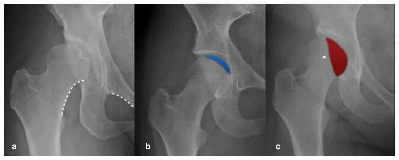
Figure 1a–c.
a) Anteroposterior (AP) radiograph of a right hip with severe lateral acetabular dysplasia, superolateral migration of the femoral head, and a break in Shenton’s line (dashed white line). b) AP radiograph of a right hip with borderline acetabular dysplasia, deficient anterior acetabular coverage, and preserved lateral center edge angle. The blue shaded area indicates the small degree of overlap between the femoral head and the anterior acetabular wall. Note the flattened sourcil or acetabular roof. c) AP radiograph of a right hip with severe lateral and posterior acetabular dysplasia. The red shaded area indicates the overlap between the femoral head and the posterior acetabular wall. Note that the center of the femoral head (white dot) lies lateral to the posterior wall, indicating a positive posterior wall sign with deficient posterior coverage.

