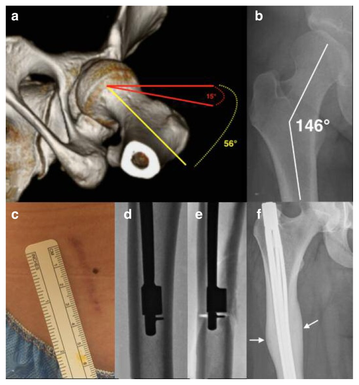
Figure 2 a–f.
a) Three-dimensional reconstruction of the left hip from computed tomography (CT) images indicating excessive femoral antetorsion of 56 degrees and resultant functional undercoverage of the anterior femoral head. b) AP radiograph of the right hip in a patient with frank acetabular dysplasia and associated excessive femoral neck shaft angle of 146 degrees corresponding with coxa valga. c) Minimally invasive surgical site used to perform a derotational femoral osteotomy (DFO) measuring 4 cm in length. d) Intraoperative fluoroscopic radiograph of the right femur with intramedullary saw in place prior to performing femoral osteotomy. e) Intraoperative fluoroscopic radiograph of the right femur with intramedullary saw in place following completion of femoral osteotomy. f) Postoperative AP radiograph of the right femur with expandable intramedullary rod in place following DFO to correct for antetorsion and coxa valga deformities. Note the well-healed femoral osteotomy site with robust callus (white arrows) and slight varus angle across osteotomy to correct for coxa valga.

