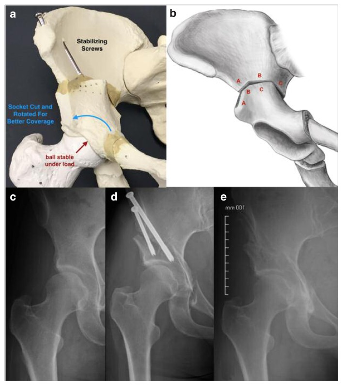
Figure 3 a–e.
a) Model representation of a periacetabular osteotomy (PAO) indicating the rotational correction of the acetabular fragment (blue arrow) which creates normalized femoroacetabular joint contact forces (red arrow) and reduces superolaterally-directed shear forces creating symptomatic instability. b) Schematic diagram of the Birmingham Interlocking Pelvic Osteotomy (BIPO) indicating location of ilium cuts (a, b, and c) and interlocking construct following rotation of central acetabular fragment. This osteotomy is stable enough for immediate full weight-bearing and, as a result, results in less muscle atrophy and deconditioning. c) AP radiograph of a right hip with borderline acetabular dysplasia predominantly due to lateral and posterior deficiencies in coverage. Note the short and inclined acetabular roof or sourcil. d) AP radiograph of a right hip following PAO with cannulated screw fixation. Note the degree of posterolateral coverage gained by the procedure with normalization of the relationship between the anterior and posterior walls. e) AP radiograph of a right hip following screw removal from healed PAO.

