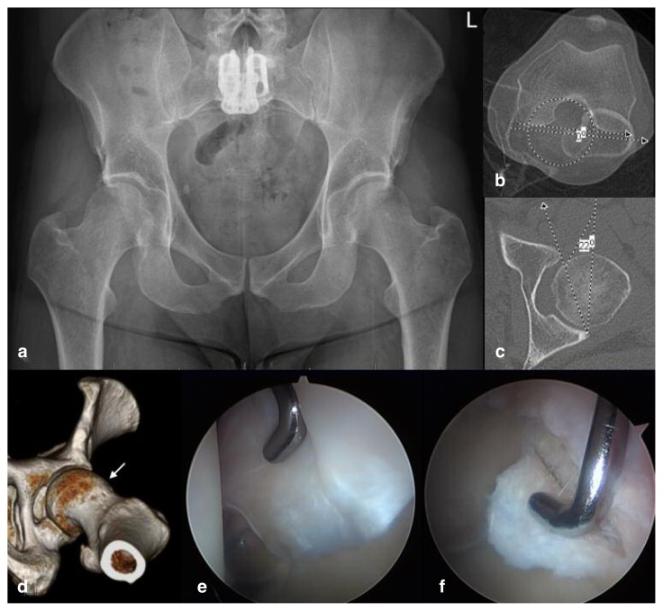
Figure 5 a–f.
a) AP radiograph of a pelvis in a 40-year-old male patient presenting with 12-month history of left groin pain and mechanical symptoms, having failed conservative management. His clinical examination is concerning for limited left hip internal rotation of 5° with the hip at 90° of flexion, despite have a severely dysplastic acetabulum. Note the positive posterior wall sign with posterior acetabular deficiency which, in conjunction with reduced lateral coverage, represents global acetabular deficiency. b) Axial CT scans of the left hip with overlapping images through the center of the femoral head, the lesser trochanter, and the distal femur. Femoral torsion measures 0° (normal 10–20° antetorsion), indicating relative retrotorsion, and accounting for the limited internal rotation seen on clinical exam. c) Axial CT scan of the left hip through the center of the femoral head indicating equatorial acetabular version to be 22° anteversion (normal 15–20° anteversion). Given the posterior wall deficiency, this value underrepresents the true extent of anterior acetabular deficiency. d) Three-dimensional CT reconstruction of the left hip indicating a large cam lesion in the upper femur (white arrow). e) Arthroscopic image of the left hip demonstrating enlarged and torn labrum consistent with instability. f) Arthroscopic image of the left hip demonstrating full thickness “outside-in” articular flap with a break in the chondrolabral junction consistent with cam-type FAI. The impingement is further exacerbated by the femoral retrotorsion. The pattern of intra-articular injury in this hip supports both a diagnosis of impingement and that of instability.

