. 2016 Nov 23;25(6):307–317. doi: 10.5607/en.2016.25.6.307

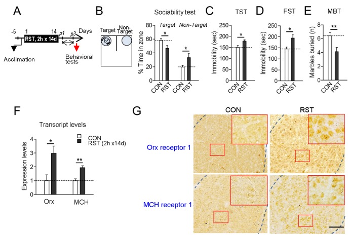
Fig. 1. Chronic restraint stress produced deficits in sociability and mood-related behaviors, and led to upregulation of orexin and MCH in the BLA. (A) Experimental design for treatment of mice with restraints for 2 h daily for 14 days (2 h×14 d RST) and subsequent behavioral tests on post-stress days 1~3 (p1~p3). Arrow: time point for tissue preparation. (B) Social interaction levels in the U-shaped field assay. The U-shaped field with a target mouse placed in a grid cage on one side and an unanimated cage on the other side is depicted (left panel). Social interaction levels were quantified by % time spent in the target and non-target zones for 5 min. The target and non-target zones correspond to the closed squares of the U-field. (C, D) Immobility time in the TST (C) and FST (D). (E) The number of marbles buried in a 30-min session of free exploration. (F) Real-time PCR data showing stressinduced up-regulation of orexin and MCH transcripts in the basolateral amygdala (BLA). n=6 animals, with 6 repeats of PCR. (G) Photomicrographs showing immunohistochemical staining of orexin receptor-1 (top panels) and MCH receptor-1 (bottom panels) in the BLA of mice treated with 2 h×14 d RST (RST) and their controls (CON). High magnifications (inset) of the squared regions (red box) in the BLA are presented. Scale bar, 200 µm. Data are presented as the mean±SEM. * and ** denote differences from the control at p<0.05 and p<0.01, respectively (for behaviors, n=7 animals for each group).

Fig. 1