. 2016 Nov 23;25(6):307–317. doi: 10.5607/en.2016.25.6.307

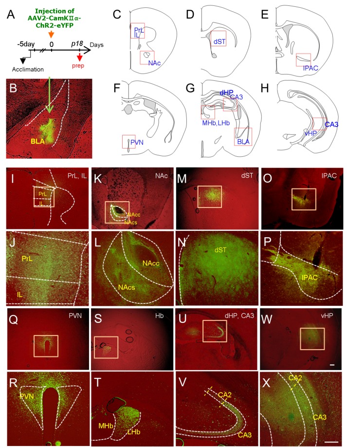
Fig. 2. Visualization of brain regions targeted by BLA neurons using the AAV2-CaMKII-ChR2-eYFP vector system. (A) Experimental design for stereotaxic injection of AAV2-CaMKIIα-ChR2-eYFP in the BLA and time point for tissue preparation (prep). AAV2-CaMKIIα-ChR2-eYFP (1.15×10¹² viral particles/ml) was injected into the right BLA, and eYFP expression was examined 18 days after injection. Six animals were analyzed using this procedure, and brains with mislocalized injections in the BLA were excluded. (B-X) eYFP fluorescence images labeling the BLA (B, injection site), medial prefrontal cortex (PrL and IL; C, I, J), nucleus accumbens, core and shell (NAc-C and NAc-Sh; C , K, L), dorsal striatum (dST; D, M, N), interstitial nucleus of the posterior limb of the anterior commissure (IPAC; E, O, P), paraventricular nucleus of the hypothalamus (PVN; F, Q, R), medial and lateral habenula (MHb, LHb; G, S, T), CA3 of the dorsal hippocampus (dHP; G, U, V), and CA3 of the ventral hippocampus (vHP; H, W, X). The areas examined and presented with eYFP fluorescence images are presented on the diagrams (C-H). Higher magnifications of squared boxes in each region are presented (J, L, N, P, R, T, V and X). Scale bars, 200 µm.

Fig. 2