. 2016 Nov 23;25(6):307–317. doi: 10.5607/en.2016.25.6.307

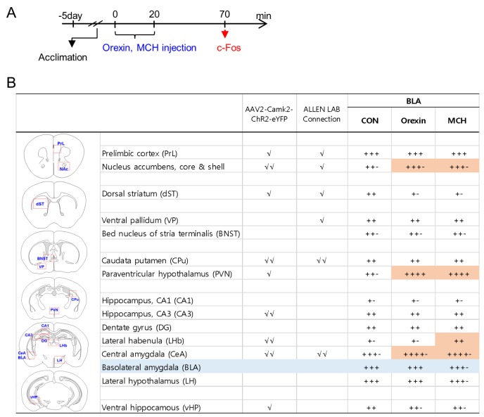
Fig. 3. c-Fos induction profiles in various brain regions following the stimulation of orexin receptors or MCH receptors in the BLA. (A) Experimental design for stereotaxic injection with orexin peptide (0.53 ng/each side) or MCH peptide (0.12 ng/each side) in the BLA, and the time point for tissue preparation (arrow). (B) Orexin- or MCH-induced c-Fos expression levels were quantified in brain sections from 6~7 animals using a 5-point rating system, and the average value for each region is presented. BLA target sites were classified as the region(s) receiving moderate (√) or extensive (√√) projections from the BLA on the basis of eYFP expression levels (AAV2-CaMKIIα-ChR2-eYFP tracking data in as seen Fig. 2, and eGFP expression levels from the Allen Lab mouse brain mapping collections (experiments #277710753 and #113144533). The areas counted for c-Fos expression in each region are marked with a red square on the diagrams. Orange colors on the table represent differences greater than an 1-scale value in c-Fos induction levels between the control and injection groups.

Fig. 3